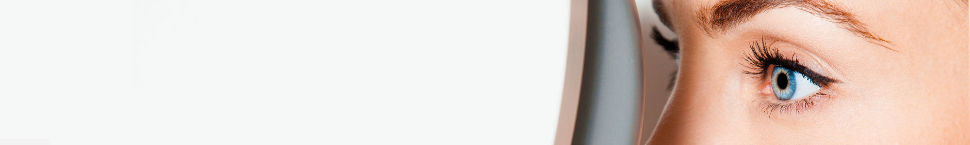
Eye Care Center has incorporated Telemedicine services in our practice. When possible, this service is utilized to reduce the spread of COVID-19. Unfortunately not all visits will qualify for Telemedicine services due to the complexity of the eye, but texting us at (979)779-9000 requesting this type of visit is what we ask all patients to do if they have an emergency need. If we determine you qualify for a Telemedicine visit, we ask that you (1) Print the consent form and email to either
Myopia, or near-sightedness, is very prevalent not only in the United States, but worldwide. Approximately 25-41% of people in the U.S. have myopia, and up to 70-90% in Asian countries! Myopia occurs when the eye is either too long, or the curvature of the front part of the eye is too strong, causing light to be focused in front of the retina. This leads to blur and difficulty with distance vision.
Corrective lenses or refractive surgical options can typically correct for this. But what about people who would like freedom from glasses during the day, but maybe are poor contact lens candidates who do not want to pursue surgical options? Is there an option for them? Or, what about athletes who need clear vision without glasses, or swimmers who can't wear contacts or glasses in the pool?! There is another option. Ortho-keratology, or Ortho-K, uses a rigid gas permeable lens, or hard contact lens, to gently flatten the cornea while you sleep. When you wake up and remove the lens, vision will be optimal for 12-15 hours OR longer without any correction! This device was approved by the FDA in 2002.
Ortho-K is also a great option for children with myopia, especially those who may benefit from myopia control. Myopia of greater than 6.00D usually indicates an increased axial length of the eye. This raises the chances of developing a retinal detachment, or other retinopathies, and even glaucoma. By slowing the progression of myopia, and the axial length of the eye, these risks can be decreased. Studies, including the SMART* study, have shown Ortho-K is a valid option for myopia control in children.
https://medcraveonline.com/AOVS/AOVS-02-00046.php
https://www.paragonvision.com/crt-lenses/
Eye Diseases: Cataracts, Diabetes, Glaucoma, Macular Degeneration
If you are diagnosed with an eye disease, you want only the best treatment available to get your eyes healthy again. At Eye Care Center, we offer only the best. Based on your diagnosis, we may recommend a wide variety of approaches, including improved nutrition, prescription medicines, therapy and vision exercises, or medical procedures / eye surgery. Ultimately, Dr. Dobson’s diagnostic skills are so vast, helping you as the patient rest better knowing you are in great hands! At Eye Care Center, we manage and/or treat dry eye, foreign bodies in the eye, eye infections (commonly known as pink eye), keratoconus, glaucoma, macular degeneration, LASIK or refractive surgery, and a host of others.
Cataracts
The natural lens in the eye gradually becomes less clear as we get older. When opacities develop in the lens and the lens gets too hazy, vision will be impaired. Our doctors can diagnose and manage cataract care. When the cataract affects the vision to the extent that you cannot read or see distant objects comfortably, we will recommend cataract surgery. Our office co-manages with cataract specialists that are proven experts in cataract surgery. Cataract surgery can provide you with a bright new world of vision.
Click here to read more about cataracts.
Diabetes
Diabetic patients should have a dilated retinal examination each year. Laser treatments have proven to slow the progression of retinal eye disease for many patients. Diabetes often stimulates the growth of new blood vessels in the back of the eye, which ultimately leak and damage the retina. If this condition is discovered early, laser treatment can destroy these vessels. We have the latest instruments used to detect changes in the back of the eye. If we discover advancing diabetic eye disease that can be treated or needs further evaluation, we will refer you for consultation and further testing to a board certified retinal specialist.
Click here to read more about diabetic retinopathy.
Glaucoma
Glaucoma gradually destroys the optic nerve tissue in the back of the eye. The greatest danger of glaucoma stems from the fact that the disease is painless and without obvious symptoms until significant damage has occurred. The most common cause is from pressure being too high inside the eye, but vascular disease and other diseases can also cause glaucoma. We always monitor for glaucoma during routine eye examinations. Tests include checking the pressure of the eye, screening for peripheral vision defects and analyzing the appearance of the optic nerve. If you are diagnosed with glaucoma, we typically prescribe medications that lower the eye pressure. Most forms of glaucoma are successfully treated with eye drops. Laser treatments and eye surgery are secondary treatments that offer alternative ways to treat more advanced glaucoma.
Click here to read more about glaucoma.
Macular Degeneration
As a disease usually associated with aging, macular degeneration is also called age-related macular degeneration (ARMD), though there are other, less common types of macular degeneration. Macular degeneration symptoms include a gradual loss of central vision needed to perform everyday tasks like driving or reading, and a reduced ability to see small visual details like fine print or patterns.
Age-related macular degeneration is the leading cause of vision loss in Americans over age 60, and presents itself in two forms: dry macular degeneration and wet macular degeneration. Of the two, the “dry” form is far more common. Both affect the center region of the retina, the light-sensitive area in the back of the eye responsible for processing images we see.
Most people with vision correction issues want to know more about LASIK and whether it is right for them. At Eye Care Center, we provide a FREE LASIK CONSULATION with our expert doctors. There is no reason to continue ‘wondering’ if you are a candidate. Schedule a free consultation today and get the facts!!
When shopping around for refractive surgeons, you must remember to “compare apples to apples,” so to speak. Surgery options can be difficult, advances in technology must be taken into consideration, and pricing gets confusing. You only get one pair of eyes, so treat them with the best. Depend on Eye Care Center doctors to provide the most ‘straight-forward’ options and choices to you, while obtaining the most accurate information.
Here is a brief description of the procedure:
A flap is surgically created in the cornea and gently folded back. An Excimer Laser is used to reshape the cornea into a flatter shape (this is similar to how a contact lens corrects vision by forming a new shape on the cornea). The flap is then put back in place, acting like a natural bandage. The healing process is quick and the discomfort level is quite low.
People will often say that they did not feel anything, and could see well the very first day. Most people have improved vision in 24-48 hours, but it must be noted that each person will heal at a different rate. Vision, although greatly improved immediately after surgery, often continues to improve for some weeks, even months.
LASIK is not for everyone. People's eyes are different so even though you may have had a friend who has had LASIK, it may not be the best course of treatment for you. If you are interested in laser vision correction, we will be happy to guide you in picking the right surgeon, procedure, and laser appropriate for you.
Laser Vision Correction Common Questions
-
What is PRK?
What is PRK?
Photorefractive Keratectomy (PRK) involves using an Excimer laser that uses a cool ultraviolet beam of light instead of a blade to reshape the cornea. It is commonly used to treat low and moderated levels of both myopia (nearsightedness) and astigmatism.
-
What is LASIK?
What is LASIK?
Laser Assisted In Situ Keratomileusis (LASIK) combines the Excimer laser with an instrument called a microkeratome. The microkeratome makes a thin flap in the superficial cornea that is lifted upwards. The Excimer laser reshapes the underlying corneal tissue, and the flap is replaced. The flap seals within 24 hours of the procedure. This procedure is used for moderate to high levels of myopia and astigmatism.
-
Are PRK and LASIK new procedures?
Are PRK and LASIK new procedures?
PRK was invented in 1987 although it was not FDA approved in the U.S. until 1995. Several million people in Europe and Canada have had this procedure prior to its approval in the U.S. The microkeratome has been in use for over ten years for other corneal procedures. It has been combined with the Excimer laser for LASIK for the last several years.
-
Are these procedures painful?
Are these procedures painful?
The procedures themselves are painless. With PRK there is discomfort after the procedure for 1 to 3 days as the epithelium (the outer corneal layer) heals. With LASIK there is little discomfort as the outer layer of the cornea is undisturbed.
-
Should I get one eye done or do both eyes simultaneously?
Should I get one eye done or do both eyes simultaneously?
Most of our patients have both eyes treated at the same time because there is only one recovery period, and the healing time is minimal. If you have only one eye treated, be aware that you may experience headaches and disorientation from the different refractions in each eye.
-
Will I be able to see better immediately?
Will I be able to see better immediately?
Vision is typically blurry immediately after either procedure. The vision clears up with PRK in approximately 1-2 weeks and within 1-2 days for LASIK. Because the healing process is different for each patient, it is hard to predict when vision will stabilize.
-
Will my vision be 20/20 after the procedure?
Will my vision be 20/20 after the procedure?
As with any procedure, we cannot guarantee perfect results after the procedure. Ten years of PRK studies show that 95% of patients see 20/40 or better without additional correction. This is legal driving vision. Some people only need their glasses for certain activities after the procedure and some have eliminated the need for glasses altogether. Presbyopia, a condition that affects EVERYONE around age 40 and requires them to wear reading glasses, cannot be corrected by any refractive surgery.
-
Is the procedure permanent or will I need to have it performed again in a few years?
Is the procedure permanent or will I need to have it performed again in a few years?
Both procedures are permanent. The tissue is completely removed and does not grow back.
-
Can I wear contacts if I still require correction after the procedure?
Can I wear contacts if I still require correction after the procedure?
Yes, the advantage to these procedures is that you can wear soft or gas permeable lenses after the procedure, if needed.
-
How soon can I have the procedure performed?
How soon can I have the procedure performed?
If you are wearing glasses and have a stable refractive error, the procedure can be performed at any time. If you are wearing soft contact lenses you will need to be out of the lenses for approximately 1-2 weeks. If you are wearing gas permeable, hard lenses, or sleep in your contact lenses, you will need to be out of the lenses for approximately 1 month.
-
Are there any restrictions on who can have the procedure?
Are there any restrictions on who can have the procedure?
We prefer to have patients treated who are over 21 years old and have a stable refraction. You are not eligible if you have diabetes, collagen vascular disease (Lupus,etc.), keratoconus, autoimmune disease (HIV), wear a pacemaker, or are a keloid former. There are also other conditions that could eliminate you as a candidate; therefore a complete eye exam is needed to determine if you are eligible.
-
When can I go back to work and when can I drive?
When can I go back to work and when can I drive?
Everyone heals at a different rate. Some people have surgery on Thursday and return to work on Friday, others take several days off work. You should not drive for at least 48 hours after either procedure.
-
Surgery Co-Management
Surgery Co-Management
Eye Care Center provides working arrangements with eye surgeons and more specialized eye care doctors to provide many of the surgical services such as cataract surgery, laser surgery, refractive surgery and strabismus surgery, retinal detachments, retinal tears and hemorrhages. You can be assured that your eyes will be comprehensively managed by the best eye care professionals.
As a fully licensed and equipped optometric practice, Eye Care Center offers a complete range of eye care services to all our patients. Whether the eye care issue involves correcting refractive errors with eyeglass or contact lenses, or helping a student find frames that will not look geeky, our experienced team will identify and implement the best eye care solutions possible.
With our years of experience in diagnosing and treating typical vision disorders such as nearsightedness, farsightedness, amblyopia, presbyopia, cataracts, macular degeneration and diabetic retinopathy, Dr. Belinda Dobson and her team are equipped and eager to provide you appropriate therapeutic medical eye care. Our practice provides complete eye care for both adults and children. Our comprehensive eye examinations will review your eyes inside and out for any potential eye disorders or diseases. Your visual skills and abilities are carefully evaluated and appropriate treatment is prescribed if needed, whether it’s medication, lenses, or vision therapy. We are prepared to handle whatever your eye care needs are, and will treat them with the latest in diagnostic equipment and technology.
There are a few items that will be needed for your eye exam. In addition to your smiling face, please bring the following to your appointment with us: Photo ID, Back-up glasses, Sunglasses, and/or Over-the-Counter Reading glasses, Medications List, and Previous Contact Lens Information, and your Vision AND Medical Insurance Cards. (Note: Our exams address the health and vision of your eyes, so in some cases your medical insurance may be applied to your visit.)
From the second you walk into Eye Care Center, our main focus is YOU. Throughout your appointment with us, you will never feel rushed or hurried. We ask that you allow approximately two hours for your appointment so you have plenty of time to experience excellence in the exam, as well as take your time selecting the most fashionable eyewear possible.
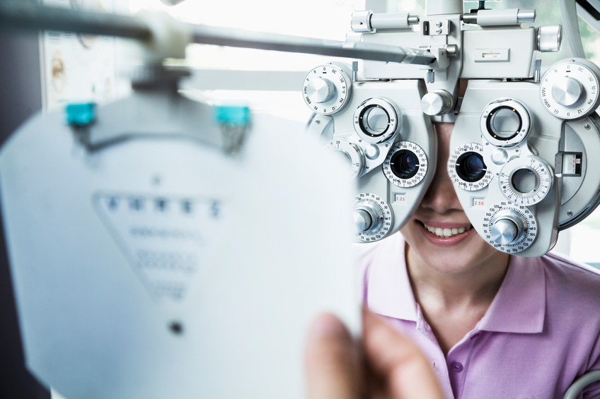
Our trained optometric technicians perform the first part of your eye exam. They efficiently gather all pertinent eye health data and vision measurements for Dr. Dobson to evaluate during your exam with her. Your personal technician will also recommend the appropriate yet optional exam upgrades, which include Optomap Retinal Photography and Peripheral Vision Screenings. Both ensure your eyes are performing up to par.
Next, Dr. Belinda Dobson proceeds with a thorough eye examination. She will assess your eyes’ visual performance, as well as the overall health. She uses only the very best technology available, and has a steadfast commitment to staying ahead of the industry in diagnostic optometry and medical equipment. From a patient’s perspective, this most often ensures a very painless exam that is chocked full of information! Dr. Dobson’s common, favorite patient compliment is hearing, “Wow, this is the best exam I’ve ever had. I had no idea what I’ve been missing.”
Additionally, during your visit at Eye Care Center, there are opportunities to view interactive educational videos on eye conditions frequently diagnosed. This allows you to be completely informed on your ocular health and visual performance.
Frequency of your eye exam is also important in prevention and the proper care of your vision and eye health. Eyecare specialists suggest that you have a complete eye exam every one to three years, depending on your age, risk factors, and physical condition.
Children
Some eyecare experts estimate that approximately 5% to 10% of pre-schoolers and 25% of school-aged children have vision issues. According to the American Optometric Association (AOA), every child should have an eye exam at 6 months of age, at age 3 and again before school starts. Children who do not have vision issues or risk factors for eye or vision problems should have their eyes checked at least every two years throughout school.
Children who have existing vision issues or risk factors need to have an eye exam more often. Common risk factors for vision issues include:
- premature birth
- developmental delays
- turned or crossed eyes
- family history of eye disease
- history of eye injury
- other physical illness or disease
The AOA suggests that children who need to wear glasses or contact lenses should have their eyes checked at least every 12 months or in accordance with their eye doctor’s instructions. Read more about Pediatric Eye Exams.
Adults
The AOA suggests that all adults who wear glasses or contact lenses have an annual eye exam. If you don’t generally need vision correction, you should have your eyes examined every two to three years until you reach 40, depending on how quickly your vision changes, and your overall health. Doctors usually recommend examinations more frequently for adults with diabetes, high blood pressure and other disorders, as many diseases can have a negative impact on vision and eye health.
When over 40, you should have your eyes examined every one or two years to check for commonplace age-related eye problems like cataracts, presbyopia, and macular degeneration. Read more about Vision After 40.
The risk of eye disease increases with advancing age, so everyone over the age of 60 should have an eye examination yearly.
Learn more about what problems can be spotted with an eye exam. Contact Eye Care Center at (979) 779-9000.
Contact lenses offer such convenience, flexibility, and versatility. Regardless of your previous experience with glasses or contacts, our eye professionals can help fit you into a pair of lenses that is just right for you. We are experts in fitting contact lenses and can handle the most specialized needs. Whether rigid, soft, scleral, colored, or clear lenses, we are experienced in handling the most difficult of prescriptions.
So you say, “But I have been told by my last eye doctor I can’t wear contacts?” While some patients are truly not candidates, Dr. Dobson’s skills and technology awareness allow her to utilize different, more “cutting-edge” contacts to “wow” those who have not tolerated contacts well in the past.
During your eye exam, Dr. Dobson will determine if you qualify for a contact lens fitting. Your glasses prescription is determined during the eye exam and is used to customize an initial contact lens prescription. We stock thousands of lenses so in most cases you leave the office the same day with your free contact lens trials. If you have never worn contacts, our trained professionals will spend quality time teaching you the proper methods for insertion and removal, as well as how to care for the contacts. When possible, we select daily contact lens. These lenses are voted the most convenient and comfortable lenses on the market. Studies prove these daily contacts are the safest contacts, and the fastest growing lens in the world.
If you have allergies, then you know allergies can be particularly annoying to contact lens wearers. In the past, many people had to quit wearing their lenses during the allergy season. However, new lens materials and allergy medications have made it possible for these people to eliminate the burning itchy eyes associated with allergies. Prescription medications such as Pazeo® or Lastacaft® offer effective and safe options, even in children as young as 3 years old. Let us know when you come in for your appointment if you have special allergy needs and we will be happy to help you.
At Eye Care Center, we have a “no strings attached” policy. This means that after you engage in the contact lens fitting process, you are not obligated to purchase contact lenses if you change your mind or are unable to wear them successfully. This allows you to try them out without purchasing contacts. The trial lenses are always free during this fitting process.
Click here to read more about our customized contact lenses.
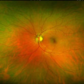
In our continued efforts to bring the most advanced technology available to our patients, Dr. Belinda Dobson is proud to announce the inclusion of the Optomap Retinal Exam as an integral part of your eye exam. The new Optomap Daytona provides the most thorough and wide-field image of the retina on the market today.
Many eye problems can develop without warning and progress with no symptoms. Early on, you might not notice any change in your vision. However, diseases such as macular degeneration, glaucoma, retinal tears or detachments, as well as other health problems such as diabetes and high blood pressure, can often be detected with a thorough exam of the retina. The retina is the part of your eye that catches the image of what you are looking at, similar to the film in a camera.
An Optomap Retinal Exam provides:
- A scan to confirm a healthy eye or detect the presence of disease.
- An overview or map of the retina, giving your eye doctor a more detailed view than he/she can achieve by other means.
- The opportunity for you to view and discuss the Optomap images of your eye with your doctor at the time of your exam.
- A permanent record for your medical file, enabling your optometrist to make important comparisons if potential problems show themselves at a future examination.
According to experts, 80% of learning is visual, which means that if your child is having difficulty seeing clearly, his or her learning can be affected. This also goes for infants who develop and learn about the world around them through their sense of sight. To ensure that your children have the visual resources they need to grow and develop normally, their eyes and vision should be checked by an eye doctor at certain stages of their development.
According to the American Optometric Association (AOA) children should have their eyes examined by an eye doctor at 6 months, 3 years, at the start of school, and then at least every 2 years following. If there are any signs that there may be a vision problem or if the child has certain risk factors (such as developmental delays, premature birth, crossed or lazy eyes, family history or previous injuries) more frequent exams are recommended. A child that wears eyeglasses or contact lenses should have his or her eyes examined yearly. Children’s eyes can change rapidly as they grow.
Eye Exams in Infants: Birth – 24 Months
A baby’s visual system develops gradually over the first few months of life. They have to learn to focus and move their eyes, and use them together as a team. The brain also needs to learn how to process the visual information from the eyes to understand and interact with the world. With the development of eyesight, comes also the foundation for motor development such as crawling, walking and hand-eye coordination.
You can ensure that your baby is reaching milestones by keeping an eye on what is happening with your infant’s development and by ensuring that you schedule a comprehensive infant eye exam at 6 months. At this exam, the eye doctor will check that the child is seeing properly and developing on track and look for conditions that could impair eye health or vision (such as strabismus(misalignment or crossing of the eyes), farsightedness, nearsightedness, or astigmatism).
Since there is a higher risk of eye and vision problems if your infant was born premature or is showing signs of developmental delay, your eye doctor may require more frequent visits to keep watch on his or her progress.
Eye Exams in Preschool Children: 2-5
The toddler and preschool age is a period where children experience drastic growth in intellectual and motor skills. During this time they will develop the fine motor skills, hand-eye coordination and perceptual abilities that will prepare them to read and write, play sports and participate in creative activities such as drawing, sculpting or building. This is all dependent upon good vision and visual processes.
This is the age when parents should be on the lookout for signs of lazy eye (amblyopia) – when one eye doesn’t see clearly, or crossed eyes (strabismus) – when one or both eyes turn inward or outward. The earlier these conditions are treated, the higher the success rate.
Parents should also be aware of any developmental delays having to do with object, number or letter recognition, color recognition or coordination, as the root of such problems can often be visual. If you notice your child squinting, rubbing his eyes frequently, sitting very close to the tv or reading material, or generally avoiding activities such as puzzles or coloring, it is worth a trip to the eye doctor.
Eye Exams in School-Aged Children: Ages 6-18
Undetected or uncorrected vision problems can cause children and teens to suffer academically, socially, athletically and personally. If your child is having trouble in school or afterschool activities there could be an underlying vision problem. Proper learning, motor development, reading, and many other skills are dependent upon not only good vision, but also the ability of your eyes to work together. Children that have problems with focusing, reading, teaming their eyes or hand-eye coordination will often experience frustration, and may exhibit behavioral problems as well. Often they don’t know that the vision they are experiencing is abnormal, so they aren’t able to express that they need help.
In addition to the symptoms written above, signs of vision problems in older children include:
- Short attention span
- Concerns for dyslexia
- Headaches
- Frequent blinking
- Avoiding reading
- Tilting the head to one side
- Losing their place often while reading
- Double vision
- Poor reading comprehension
The Eye Exam
In addition to basic visual acuity (distance and near vision) an eye exam may assess the following visual skills that are required for learning and mobility:
- Binocular vision: how the eyes work together as a team
- Focusing
- Peripheral Vision
- Color Vision
- Hand-eye Coordination
- Tracking
The doctor will also examine the area around the eye and inside the eye to check for any eye diseases or health conditions. Most times, this requires a dilated pupil exam, which opens the pupil to (1) allow Dr. Belinda Dobson to examine the retina and internal eye structures, as well as (2) relax the eye muscles to determine the amount of focus the child focuses on average. You should tell the doctor any relevant personal history of your child such as a premature birth, developmental delays, family history of eye problems, eye injuries or medications the child is taking. This would also be the time to address any concerns or issues your child has that might indicate a vision problem.
If Dr. Belinda Dobson does determine that your child has a vision problem, she may discuss a number of therapeutic options such as eyeglasses or contact lenses, an eye patch, vision therapy, depending on the condition and the doctor’s specialty. Since some conditions are much easier to treat when they are caught early while the eyes are still developing, it is important to diagnose any eye and vision issues as early as possible.
Following the guidelines for children’s eye exams and staying alert to any signs of vision problems can help your child to reach his or her potential.
 At Eye Care Center, we strive to exceed your expectations. It is top priority that our patients receive the utmost level of care, feel comfortable and ‘at ease’ during the exam process, and overall leave more educated about their eyes than before their appointment.
Sometimes ‘fear of the unknown’ can hinder this process, especially for children, special needs patients, and those going to the doctor for the first time. We strive to minimize any anticipation, anxiousness, and aversion toward going to the doctor and instead want it to be fun and interactive. With this in mind, we “think outside the box” and offer an alternative to the traditional eye exam. Laci is our Golden-Doodle dog who we use as a full-time, on-site therapy or comfort dog. She is very popular with our patients, and is great at calming nerves and easing anxiety. Studies show that even a short-term exposure to a comfort dog has beneficial physiological and psychosocial effects on the patients who want it.
And remember, Laci is not just for the young patients. We could all use some “Laci Loving” from time to time. Don’t forget to request to see her at your next appointment.
Expert treatment for pink eye, eye infections and foreign object removal
In the event of an eye emergency, you don’t always need to rush to the emergency room or primary care physician. At Eye Care Center, our optometrist is qualified and experienced to treat many eye emergencies.
With advanced optometric technology in our comfortable, customer service driven clinic, we’ll help resolve any condition(s) that pose danger to your eyes and/or vision. Visit us when you have an eye emergency and you’ll receive effective treatment that’s also convenient and cost-effective.
Eye Symptoms that Require Immediate Medical Attention
Many eye and visual complaints require immediate care, and it’s critical not to delay treatment for these eye emergencies. Contact us promptly if you experience any of the following:
- A foreign object scratches or gets lodged in your eye
- Trauma to your eye, such as exposure to a harmful foreign substance or burns
- When new “floaters” appear in your vision
- Sudden flashes of light
- Soreness, itchiness, pain, redness and/or oozing, such as in “pink eye”
- Ocular allergic reactions
- Sudden loss of vision, in either one or both eyes
- When a contact lens breaks or becomes dislodged in your eye
Our professional eye doctor, Dr. Belinda Dobson O.D. will examine your eyes skillfully with the latest diagnostic tools. She will inspect your cornea thoroughly, and if necessary she’ll perform a digital scan of your inner eye to assess for any possible injuries or damage.
Pink Eye
Formally known as conjunctivitis, pink eye refers to an infection of the membrane (conjunctiva) that covers the white part of your eye and lines your eyelid. The characteristic reddish or pink eye color comes from an inflammation of the small blood vessels in your conjunctiva.
A bacterial or viral infection, or an allergic reaction, is usually to blame for pink eye. While this condition is annoying, it usually has no effect on vision. It may be contagious though, so early detection and treatment is important. Contact us to schedule an appointment today in our Bryan/College Station office as soon as possible.
Pink Eye? What to do in the meantime, before your optometrist appointment
If you wear contact lenses, remove them immediately and don’t wear them as long as you have symptoms of pink eye. Take care to wash your hands well and frequently, and don’t share towels – this goes far towards preventing the condition from spreading to those around you.
Applying cold or warm compresses to your eyes for a few minutes, a few times a day, may help relieve your symptoms until you reach your eye doctor.
Foreign Object Removal
This is one of the most common types of eye emergencies. Symptoms include pain, irritation and redness. Regardless of whether the offending object is an eyelash, sand or a splinter of wood, your body will first react with an inflammatory response that causes swelling. Sometimes you will be able to rinse the object out of your eye successfully, and the discomfort will be alleviated. However, if this doesn’t work, it’s time to contact us for help from an eye care professional. Using specialized instruments or an effective eye wash, we will remove the foreign object gently, thereby preventing further damage to your delicate eye tissues.
NOTE: If you are suffering from extreme pain, it’s advised to go to your local hospital emergency room for immediate care. This may be a sign that the foreign object has penetrated the outer layer of your eye, which can cause vision loss or retinal damage.
24-hour Emergency Care
Eye injuries range from the very minor such as getting soap in your eye, to the catastrophic such as chemical exposures or lacerations, which could result in permanent loss of vision. Eye Care Center understands the importance of immediate eye care when there are symptoms of sudden vision loss, pain, light sensitivity, discharge, redness, flashes of light, floaters, etc. These are signs that can require an immediate evaluation or consultation. Our office provides emergency services for eye infections and eye injuries. Please call our office at (979) 779-9000 during office hours or for emergencies. If outside normal business hours, you will be prompted through an automated system that will help you contact our physician on call. In most cases, Dr. Belinda Dobson optometrist will be able to return your call but if not, please seek medical attention at the hospital / urgent care or dial 911.
Our staff will work with you to schedule an appointment as soon as possible and walk-in’s are always welcome. State of the art microscopes allow us to examine the front surface of the eye and facial areas around the eye for infection or injury. After assessing the extent of the injury or infection, a treatment plan will be formulated and explained to you. Treatment may include medications and supportive care. Follow-up visits to monitor your recovery will be scheduled as needed.
Come to Eye Care Center in College Station for eye emergency treatments.
Dry Eye
Dry eye is quite perhaps one of the most under-diagnosed diseases in our country. At Eye Care Center, our passion is identifying patients with dry eye, educating them on the disease process, and formulating a proper eyecare regimen to eliminate their signs and symptoms.
Symptoms of Dry Eye:
- Blurry vision
- Fluctuations in vision
- Burning & stinging
- Gritty eyes
- Foreign body sensation
- Watering
- Itchy eyes
- Light sensitivity
- Painful eyes
If you have any of these symptoms, timely intervention and treatment is critical. We offer dry eye treatment in Bryan / College Station at Eye Care Center. As your optometrist, we are experts in examining your eyes for dry eye syndrome and treating them in the best way possible. We offer treatments for dry eyes ranging from prescription eye drops to punctal plugs and everything in between. Have our eye doctor examine your eyes to determine the best dry eye treatment for them.
Dry eyes or dry eye syndrome (DES) is an ongoing condition that treatments may be unable to cure. But the symptoms of dry eye—including dryness, scratchiness and burning—can usually be successfully managed. Artificial tears, which are lubrication eye drops may alleviate the dry, scratching feeling and foreign body sensation of dry eye. We are also fortunate to now have prescription eye drops. Because there are three layers of the tear film, it is important to understand which layer of tear film is the cause of dry eye. Each layer can require a different treatment approach. Some work on each layer of the tear film, some work on increasing production and some work on improving the quality of the tears.
Check with Dr. Belinda Dobson before buying any over-the-counter eye drops. Many over the counter eye drops have harsh preservatives or chemicals that can be toxic to the cornea, and can actually worsen your dry eye.
To reduce the effects of sun, wind and dust on dry eyes, wear sunglasses when outdoors. Wraparound styles offer the best protection.
Indoors, an air cleaner can filter out dust and other particles from the air, while a humidifier adds moisture to air that's too dry because of air conditioning or heating.
Bryan / College Station Dry Eye Treatment for Severe Cases
For more significant cases of dry eye, Dr. Dobson may recommend punctal plugs. These tiny devices are inserted in ducts in your lids to slow the drainage of tears away from your eyes, thereby keeping your eyes more moist. In addition, many times our doctors recommend Omega 3 Fatty Acids which have been proven to help with Dry Eye Syndrome. There are also new break through medications that are revolutionizing dry eye. Schedule your appointment today with Dr. Dobson to see if you are a candidate for this dry eye therapy.
If medications are the cause of dry eyes, discontinuing the drug generally resolves the problem. But in this case, the benefits of the drug must be weighed against the side effect of dry eyes. Sometimes switching to a different type of medication alleviates the dry eye symptoms while keeping the needed treatment. In any case, never switch or discontinue your medications without consulting with your doctor first.
Treating any underlying eyelid disease, such as blepharitis, helps as well. This may call for antibiotic or steroid drops, plus frequent eyelid scrubs with an antibacterial shampoo. If you are considering LASIK, be aware that dry eyes may disqualify you for the surgery, at least until your dry eye condition is successfully treated. Dry eyes increase your risk for poor healing after LASIK, so most surgeons will want to treat the dry eyes first, to ensure a good LASIK outcome. This goes for other types of vision correction surgery, as well.
Ocular Allergies
While the Brazos Valley & Bryan/College Station are definitely lovely places to live, the allergies that come along with them leave some feeling aggravated and uncomfortable almost year round. If you suffer from allergies and its’ effects on the eyes, let Dr. Belinda Dobson help you today.
Eye allergy occurs when something you are allergic to irritates the conjunctiva, which is a delicate membrane covering the eye and the inside of the eyelid. Like all allergies, allergic conjunctivitis starts when the immune system identifies an otherwise harmless substance as an allergen. Symptoms can onset within minutes to hours in more acute cases, yet linger for months in more chronic cases.
Symptoms of Ocular Allergies:
- Red eyes
- Watering eyes
- Itching or burning
- Sensitivity to Light
- Gritty eyes
- Puffy or swollen eyes or lids
The most common causes of allergic conjunctivitis are seasonal allergens such as pollen and mold spores. People with seasonal allergic rhinitis (hay fever) normally notice their symptoms worsen when they go outdoors on days with high pollen counts.
Indoor allergens such as dust mites and pet dander can also cause eye allergies year-round. If you suffer from this type of allergy, you may notice your symptoms worsen during certain activities such as cleaning your house or grooming a pet.
Eye allergy symptoms can be very annoying. Yet they pose little threat to eyesight other than temporary blurriness. Unlike conditions such as pink eye, allergic conjunctivitis is not contagious. The first step toward relief is proper diagnosis. Dr. Belinda Dobson will use her high power microscopes to examine the anterior surface of the eye and inspect for signs of allergic conjunctivitis.
After proper eye allergy diagnosis, she will provide a regimen to eliminate your symptoms quickly, and prevent future episodes of allergic conjunctivitis.
If you suspect you suffer from ocular allergies, wait no longer. Schedule your appointment with Eye Care Center and let our eyecare professionals help you find the road to relief quickly.
Keratoconus is a degenerative or progressive disorder that affects the cornea. The cornea is the clear membrane that covers the colored part of the eye and pupil. The cornea is the “window” of the eye and is the most powerful lens in the eye as well. Keratoconus is a corneal disease that causes structural changes within the cornea which cause the cornea to thin and bulge outward into a steeper, irregular, more conical shape than its normal gradual curve.
Keratoconus can cause substantial visual loss of vision, image distortion, streaking of lights, sensitivity to light, and multiple images, etc. Keratoconus affects approximately one person in a thousand. However, the exact cause of keratoconus is uncertain. It has been linked to genetic factors and associated with detrimental abnormal enzyme activity in the cornea. However, the findings are still inconclusive.
Many patients with keratoconus may be treated with corrective lenses, glasses, contact lenses, intrastromal corneal ring segments, and as a last resort, corneal transplantation. In order to stabilize the cornea, keep the keratoconus from progressing and even avoid having to have a corneal transplant, many patients now have chosen to travel around the world to have corneal collagen crosslinking, also known as "CXL."
At Eye Care Center, we proudly announce that we work closely with the world-class experts in the field of keratoconus. At our office in College Station, we maintain extensive diagnostic technology to diagnose and track the progression of keratoconus. When surgical intervention is needed, we work with our colleagues in Houston at Slade & Baker Vision Center. As leaders in the field, Slade & Baker were the first FDA Approved site for clinical trial in Houston, Texas and are still currently involved in this study. Our patients in the Bryan / College Station participated in this cutting edge study and are still seeing excellent results today.
If you or someone you know has been diagnosed with keratoconus and you live in or near the Houston area, please feel free to contact us for more information. We encourage you to visit our Crosslinking page for more information on this procedure and its wonderful results.
Contact lenses are fit for many reasons including correcting nearsightedness, farsightedness, astigmatism, and presbyopia. They are also used for therapeutic purposes such as after refractive surgery or for keratoconus. Athletes, young patients who are not yet ready for refractive surgery, and patients who need bifocals but do not wish to wear glasses can also benefit from wearing contact lenses. In many of these patients, vision is actually better in contact lenses rather than glasses / spectacles. Below are several types of contacts we recommend and use daily on our special fit patients.
If you've been told in the past that you cannot wear contact lenses because of an irregular cornea or other problems, you may want to get a second opinion and ask your eye doctor about scleral contact lenses.
Scleral contacts are large-diameter gas permeable contact lenses specially designed to vault over the entire corneal surface and rest on the "white" of the eye (sclera). In doing so, scleral lenses functionally replace the irregular cornea with a perfectly smooth optical surface to correct vision problems caused by keratoconus and other corneal irregularities.
Also, the space between the cornea and the back surface of a scleral lens acts as a fluid reservoir to provide comfort for people with severe dry eyes who otherwise could not tolerate contact lens wear.
Many optometrists recommend scleral contact lenses for a variety of hard-to-fit eyes, including eyes with keratoconus. Scleral contacts are noticeably larger than standard gas permeable (GP) contacts and have a diameter equal to or greater than that of soft contact lenses. The smallest sclerals are approximately 14.5 mm in diameter, and the largest can be up to 24 mm. When most people think about putting a large contact in their eyes, they assume the comfort will be poor. With our patients at Eye Care Center, we have found the response to be quite the opposite. The lenses provided excellent comfort.
In cases of early keratoconus, a standard GP lens may be used. However, if the lens does not center properly on the eye or moves excessively with blinks causing discomfort, switching to a large-diameter scleral contact lens may solve the problem.
Because scleral lenses are designed to vault the corneal surface and rest on the less sensitive surface of the sclera, these lenses often are more comfortable for a person with keratoconus. Also, scleral lenses are designed to fit with little or no lens movement during blinks, making them more stable on the eye, compared with traditional corneal gas permeable lenses.
If you or someone you know is interested in trying scleral lenses for keratoconus or other ‘hard-to’fit’ eyes, please contact us at (979) 779-9000 or schedule an appointment today.
If you have keratoconus or another irregular cornea condition, traditional contact lenses can sometimes be problematic. Many people experience limited wear time, discomfort after a few hours of wear or glare from their current contact lenses. Most of the traditional lenses do not allow for good, stable vision and leave the patient frustrated (and blurry)!
With KeraSoft® IC there is an alternative. Its innovative design uses the latest technologies to create a soft contact lens that fits many unusually shaped corneas – one you can wear comfortably. Dr. Belinda Dobson enjoys the benefits of the lens with her patients because it is customized to specific corneal shape and prescription needs, its designed to provide excellent comfort and vision, and ensures a high, healthy level of oxygen.
Gas permeable contact lenses are rigid lenses made of durable plastic that transmits oxygen. These lenses also are called GP lenses, rigid gas permeable lenses, RGP lenses and oxygen permeable lenses. For many patients, they offer outstanding benefits over soft lenses. For one, because GP lenses are made from a firm plastic material, they retain their shape when you blink, which tends to provide sharper vision than pliable soft lenses.
GP lenses also are extremely durable. Although you can break them (for instance, if you step on them), you can't tear them easily, like soft lenses. In addition, they are made of materials that don't contain water (as soft contact lenses do), so protein and lipids from your tears do not adhere to GP lenses as readily as they do to soft lenses.
There are some disadvantages of RGP lenses for patients with irregular corneas and most of these deal with discomfort and poor fit. Because of the shape and curvature of more keratonic corneas, the RGP lens has a difficult time centering, thus creating movement of the lens, and therefore blurring vision and making the lens uncomfortable.
Corneal Crosslinking offers a solution for slowing or halting the progression of keratoconus. For patients who have keratoconus, crosslinking provides an attractive and minimally invasive tactical approach to slow the disease. At Eye Care Center, Dr. Belinda Dobson examines the cornea and determines if one is a candidate for crosslinking. If we determine corneal collagen crosslinking is an option for the management of keratoconus, we perform the pre-operative examination and carefully work with Dr. Slade’s office to orchestrate the treatment phase.
Purpose:
Corneal Crosslinking (CXL) is an investigational treatment for Keratoconus patients and Post Lasik Ectasia patients that experience corneal thinning after Refractive Surgery. This trial is studying if this one-time treatment will strengthen the cornea so that the progression of Keratoconus and Corneal Ectasia is slowed or stopped.
Background:
Corneal Collagen Crosslinking (CXL) has been proven in studies outside of the US to strengthen a weakened corneal structure, as in keratoconus. CXL is currently in US Food and Drug Administration (FDA) clinical trials to seek FDA approval.
Keratoconus/Effects:
The cornea is the clear membrane that covers the colored part of the eye and pupil. The cornea is the “window” of the eye and is the most powerful lens in the eye as well. Keratoconus is a corneal disease that causes structural changes within the cornea causing the cornea to thin and bulge outward into a steeper, irregular, more conical shape than its normal gradual curve. Keratoconus can cause substantial visual loss of vision, image distortion, streaking of lights, sensitivity to light, and multiple images, etc.
Keratoconus affects about one person in a thousand, and yet the exact cause of it is uncertain. It has been associated with genetic factors and linked to detrimental abnormal enzyme activity in the cornea; however, the findings are still inconclusive. Many patients with keratoconus may be treated with corrective lenses, glasses, contact lenses, intrastromal corneal ring segments, and as a last resort, corneal transplantation. In order to stabilize the cornea, keep the keratoconus from progressing and even avoid having to have a corneal transplant, many patients now have chosen to travel around the world to have corneal collagen crosslinking, also known as "CXL."
Goal:
Crosslinking (CXL) has been shown to increase the number of corneal crosslinks within the cornea. These links are like the natural anchors in the cornea and are responsible for preventing the cornea from bulging outwards and becoming steep and irregular. The goals of crosslinking are to stop the progression of keratoconus, decrease the severity of the corneal bulging, and allow the patient to continue or resume contact lens wear. By decreasing the severity of the corneal bulging, doctors are better able to fit the patient for contact lenses.
Study Parameters:
You may be eligible for this study if you are 12 years of age or older and have been diagnosed with Keratoconus or diagnosed with Corneal Ectasia after refractive surgery (e.g. LASIK, PRK). There are specific inclusion and exclusion criteria that patients must meet in order to be considered participants in the clinical trial. We will be happy to discuss these with you.
Procedure:
The crosslinking treatment is an outpatient procedure performed in the surgical laser suite using numbing eye drops. During this study, the surgeon uses an ultraviolet-A illumination device (UVA light treatment), called the KXL system in combination with VibeX (vitamin B2) eye drops to treat the cornea (front of the eye). First, in the Epithelial Off procedure, the outer layer of the cornea, the epithelium, is prepared for the procedure. If a patient is having the Epithelial On procedure, this step is skipped. Next, vitamin B2 (riboflavin) eye drops are instilled in the eye and the patient is asked to look at an ultraviolet light while lying comfortably in a reclining chair. The entire procedure generally takes only a few minutes. The patient’s eyes are numbed through the use of anesthetic drops.
After treatment:
Post operatively, most patients start to notice the effects in their vision 4-8 weeks after the traditional (epithelial-off) procedure and the final effects usually take 3-6 months. Every patient is different, visual results may differ.
If you are interested in learning more about CXL or keratoconus, please call us at (979) 779-9000 and ask for Madeline.
Please visit the National Keratoconus Foundation's website to see all the approved CXL sites in the United States.
If you or someone you know has been diagnosed with cataracts, you should know that this is a common condition and treatment options are available. Just because someone has cataracts does not mean they will lose vision or suffer long term.
Cataracts are a disease of the eye that results in the clouding of the lens of the eyeball, which is located directly behind the pupil. This lens acts as a “window” for your vision. Cataracts prevent clear images from appearing on the eye’s retina; causing mild, moderate, even severe blurred vision.
Typically an eye disorder associated with aging (over half of the people in America over age 80 have either had a cataract or cataract surgery), cataracts generally occur later in life as the lens structure within the human eye changes and gets older. In diabetic patients or patients with more exposure to UV light over the years, cataracts can onset at a much earlier age.
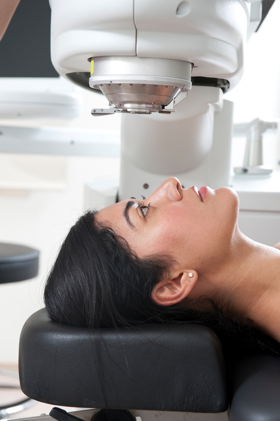
Bladeless Laser Cataract Surgery is now a reality thanks to the pioneering work of Dr. Stephen Slade and his partner Dr. Richard Baker. Dr. Dobson works very closely with Drs. Slade and Baker. On February 26, 2010, Dr. Slade performed the first cataract surgery in the United States using a femtosecond laser. LenSx Lasers, Inc. received the first laser clearance for cataract surgery by the FDA and selected Dr. Slade to perform the first procedure. Bladeless Laser Cataract Surgery uses the femtosecond laser to do many of the steps currently performed by hand, and is designed to provide a greater level of precision and safety to modern cataract surgery.
Watch a quick video on Laser Cataract Surgery:
“I have been involved in many new technology introductions, and I know from these past experiences that Bladeless Laser Cataract Surgery, will be widely accepted by surgeons and demanded by patients all over the world,” said Dr. Slade after the procedure. “This is the cataract surgery that I would want for my friends, my family and myself.”
If you decide to upgrade the basic laser cataract surgery to laser cataract surgery, Dr. Dobson co-manages patient care with Dr. Stephen Slade locally. The surgery itself is performed in Slade & Bakers “state-of-the-art” surgical center, utilizing only the most advanced technology, but Dr. Dobson performs postoperative visits locally.
Intraocular lenses (IOL) are implantable lenses that are inserted into the eye during cataract surgery. These lenses are made of plastic or silicone materials and placed permanently in the eye after removal of the eye’s natural lens. Intraocular lenses may be either monofocal or multifocal. Monofocal lenses have a single zone of clear focus, usually set for excellent distance vision, but require the use of reading glasses for near tasks, like reading or sewing. Multifocal lenses have several zones of clear vision and allow for both distance and near vision correction.
At Dr. Belinda Dobson, we know that living life to the fullest means being able to see your world clearly. As we age, our ability to maintain focus begins to decrease and our normal lens becomes cloudy and opaque. We offer premium lens implant options for after cataract surgery or before cataract development with a procedure known as Refractive Lens Exchange (RLE).
One of the most exciting advances in premium lens implant options is the Crystalens™. If you are having trouble seeing the golf ball after a long drive or simply having trouble reading, perhaps you should discuss your options with your ophthalmologist to determine if a premium lens could help you see your world in full clarity. Certain qualified patients over the age of 50 who are myopic or hyperopic can receive Crystalens™ as part of a clear lens exchange procedure also called refractive lens exchange. Crystalens™ provides the opportunity for someone to live your life without being dependant on glasses or contact lenses. If you are interested in finding out if you are a candidate please feel free to start the process by completing our self-evaluation test.
We are often asked if cataract surgery can “fix” astigmatism. The answer is absolutely! Many options are available for this type of correction. With Toric IOL - AcrySof® Toric / Astigmatic Premium Lens Implants, you can think of cataract surgery as refractive surgery as well. If you are seeking a cataract surgery expert and have astigmatism please feel free to consult with Dr. Belinda Dobson at Eye Care Center. There are NEW options available for patients that have astigmatism and need an IOL for after cataract removal. The AcrySof® Toric IOLs are artificial lenses which are ideal for individuals who have cataracts and also suffer from astigmatism. This lens is manufactured by Alcon Technologies a widely known and respected producer of intraocular implants.